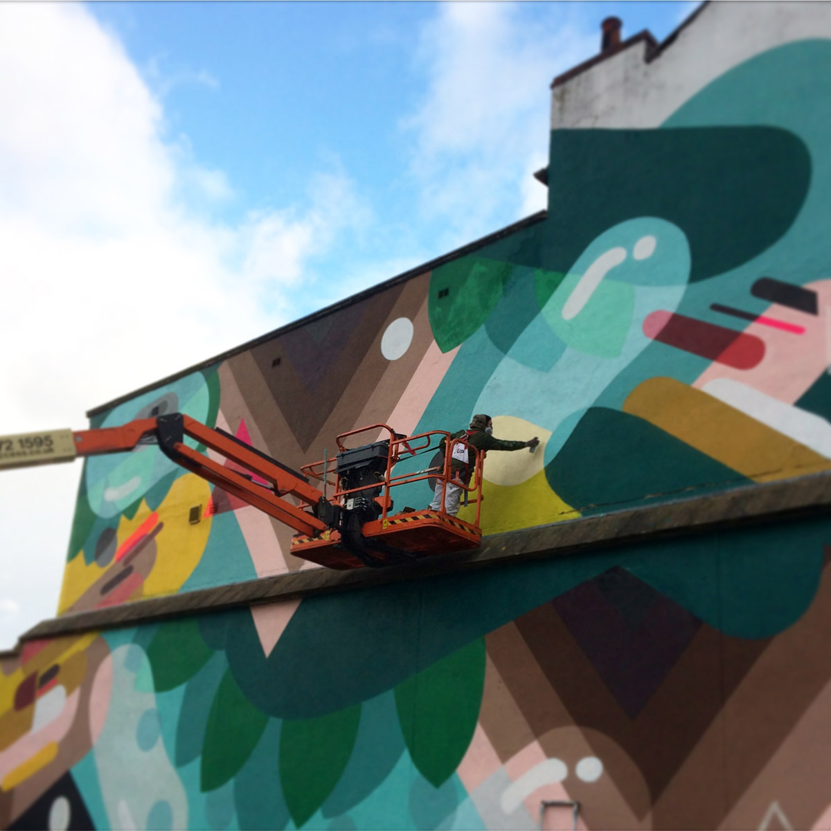
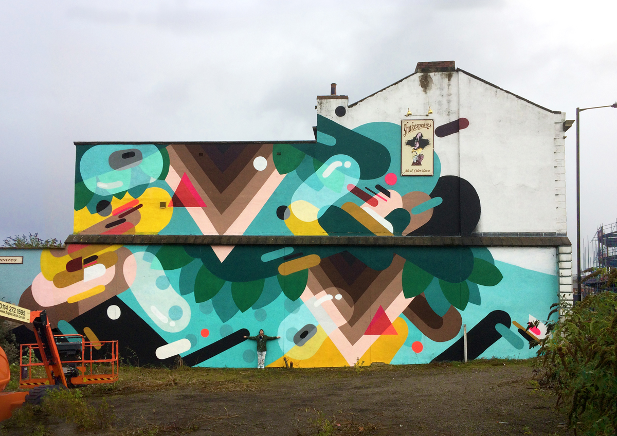
New mural on Holy Green, Sheffield UK

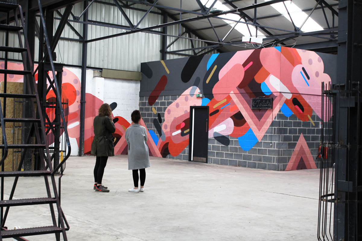
At the edge of Holy Green, where classrooms once stood and chalk dust once hung in the air, a mural blooms across the wall – a burst of colour, pattern, and quiet intent. Titled ‘The Equation for Freedom, Peace, and Equality’, it borrows from the French national motto, ‘Liberté, Égalité, Fraternité’, but leaves behind the weight of old hierarchies, exchanging “brotherhood” for a softer, more open word: peace.
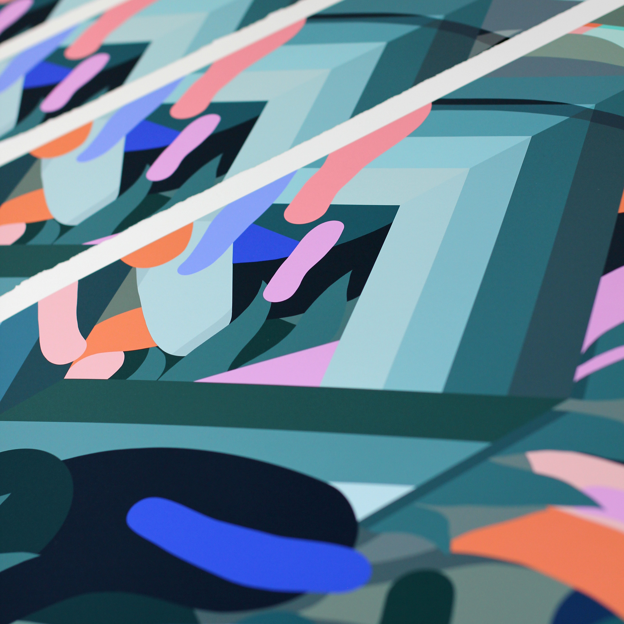
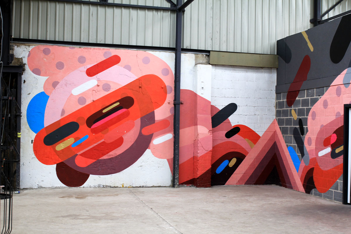
The mural pulses with energy. Bright blues, deep reds, flashes of yellow and violet woven into geometric forms that seem at once organic and mathematical. There are echoes of molecules, of cells under a microscope, of unseen worlds made visible. These patterns speak to the artist’s background in science: molecular biology, microscopy, where beauty often hides in the smallest things.
But this is not a scientific diagram. It’s a kind of visual poem. Shapes flow into each other like ideas in conversation. Overlapping, evolving, never fixed. Like the values it names freedom, peace, equality, it resists simple answers.
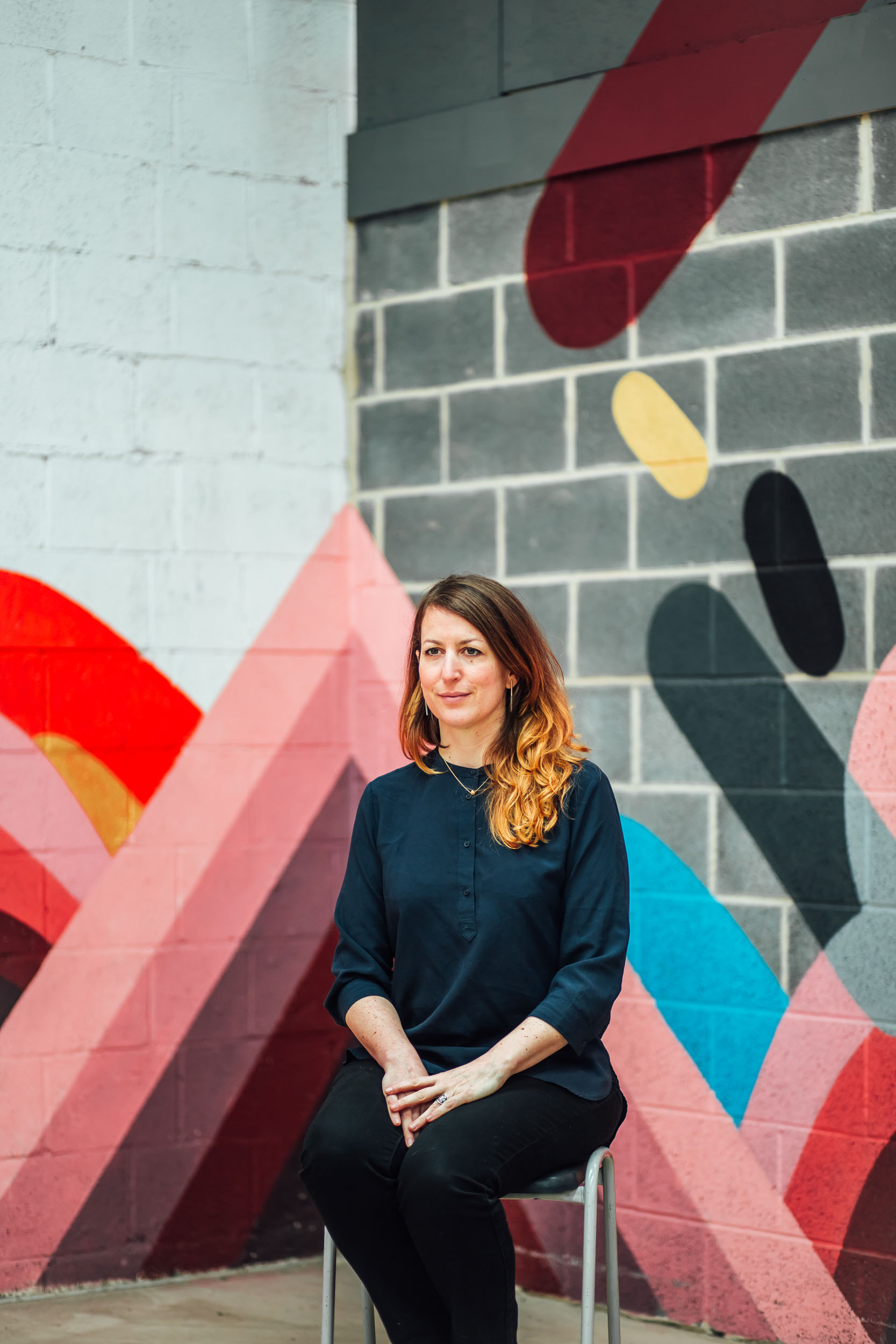
Holy Green holds its own layers of memory. Once a place of learning, it now hosts this bold, quiet call to reflect, to question. There’s a whisper of a former French teacher here, eccentric and unforgettable, maybe still echoing in the walls. A shared nationality with the artist adds one more thread to the tapestry.
If there is an equation in this mural, it’s unfinished. Drafted in hope, not certainty, this work sketches the outline of a better world. And perhaps, just perhaps, if we can solve it and truly understand the balance it hints at, we might unlock the deeper formula for peace, freedom, and equality in the universe.
It doesn’t offer solutions. Just a space to imagine them.


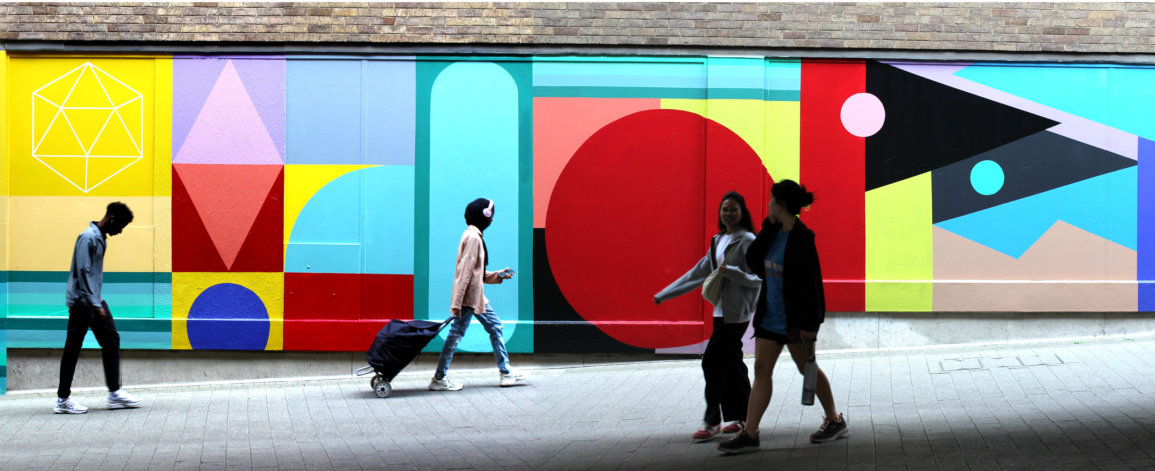

THE EQUATION FOR FREEDOM, PEACE AND EQUALITY was funded by Atkinsons, Sheffield City council and produced with the help of Friends Of Sheffield City Centre. Photos and text © Florence Blanchard 2025